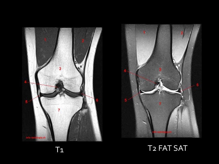
MRI is the most useful exam because it establishes the location, extent and benign characteristics of the tumor. Treatment consisted of regional synovectomy by open surgery or arthroscopy in 66.7 and 15.6% of cases, respectively.Ĭonclusions: KSH should be considered in the differential diagnosis of adult patients with chronic low-intensity knee pain. MRI was the most commonly used imaging test (98%). Pain, swelling and haemarthrosis were frequently reported (88.2, 66.7, and 47.1%). The bony tissue of the knee was rarely affected (13.5%). The suprapatellar and patella-femoral joint compartment was the most frequent location (47.9%). A literature review showed that KSH appears most frequently in children and teenagers (64.6%) and does not differ by gender. A pain-free knee without recurrence was achieve in all cases except one, which was successfully reoperated.

Histological type was arteriovenous in three cases and capillary in one.

Open excisional biopsy and regional synovectomy were performed in three patients, and by arthroscopy of the posterior compartment in the fourth. MRI revealed a benign tumor mass in all cases except one. Persistent anterior knee pain was the main complain. Three lesions located in the suprapatellar pouch, two eroding the patella and one the supratrochlear bone, and one in the posterior compartment. Results: Four adults (20–40 years) were diagnosed with KSH. A literature search was conducted in PubMed from 2000/01 to 2020/06 using the search terms “synovial haemangioma” and “knee.” Fifty full-text articles that included a total of 92 patients were included for further discussion. open resection, and follow-up in the literature.ĭesign: From 1996 to 2016, four patients with KSH were retrospectively reviewed. All patients were evaluated by 3 and 1.5 Tesla MRI units.Objective: The aim was to report 4 patients with intra-articular knee synovial haemangioma (KSH) and to perform a systematic review to describe the patient characteristics, patterns of tumor location, clinical presentation, usefulness of imaging examinations, pros and cons of arthroscopic vs. The aim of this article is to review muscle tears of unusual location, particularly in the pelvic area, but also evaluating the chest wall, abdomen, and upper and lower limbs. Magnetic resonance imaging (MRI), with a better anatomical resolution and multiplanar capability, is the method of choice for detecting the precise location and severity of the injury and can establish their severity. However, there are a significant number of muscle injuries, considered uncommon, that may be not be detected by ultrasound, mainly because of their depth, and could be responsible for long periods of inactivity for the sportsman. The diagnosis is easily made with an ultrasound study. Current studies show that 30% of injuries in athletes affect muscles, with hamstrings, quadriceps, gastrocnemius, and adductors being particularly prevalent. Muscle injuries are currently particularly frequent among people who participate in sports. En todos los casos, se usaron equipos de alto campo 1.5 y 3 Tesla.

La resonancia magnética (RM), por su resolución anatómica y capacidad multiplanar, es el método de elección para el estudio de este tipo de afecciones, ya que permite descartar otras patologías de similar presentación clínica y realizar un diagnóstico específico.Įn este artículo describimos los desgarros musculares de localización inusual, particularmente los de localización pelviana, evaluando también la pared torácica, abdominal y miembros superiores e inferiores. Sin embargo, existe un número importante de lesiones musculares de localización profunda e infrecuente, que pueden pasar inadvertidas en la ecografía y que causan largos períodos de inactividad para el deportista. El diagnóstico se realiza en forma sencilla mediante un estudio ecográfico. Según los estudios actuales, un 30% de las lesiones en atletas afecta los músculos, siendo particularmente comunes a nivel de los isquiotibiales, el recto anterior de los cuádriceps, los gemelos y los aductores. En el presente los desgarros musculares son una causa muy frecuente de lesión en la práctica deportiva.


 0 kommentar(er)
